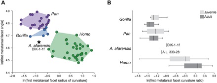
Fig. 3. Medial cuneiform ontogeny in apes, humans, and A. afarensis.

(A) The radius of curvature of the distal medial cuneiform facet and the angulation of the facet relative to the navicular facet are plotted for juvenile humans (green), gorillas (blue), and chimpanzees (purple). Humans have distally directed facets (angle ~90° to 100°), with facet convexity that ranges from ape-like in small juveniles (see fig. S5) to flatter joints in subadults (9). Ape juveniles have convex, medially directed facets. The DIK-1-1f morphology is intermediate between humans and gorillas, with a facet orientation between human and ape, and a more ape-like joint convexity. (B) When standardized by the dorsoplantar height of the medial cuneiform, facet curvature can be assessed independent of size. While facet curvature differs little between juvenile and adult apes, humans experience a developmental flattening of the joint with growth. Both DIK-1-1f and A.L. 333-28 are more convex than similarly sized humans but less so than apes and, when paired, have the ape-like pattern of maintained convexity with growth of the bone (fig. S5). The box-and-whiskers plot shows the mean (dark vertical line), upper and lower quartiles (boxes), and range (whiskers).
