Fig. 6.
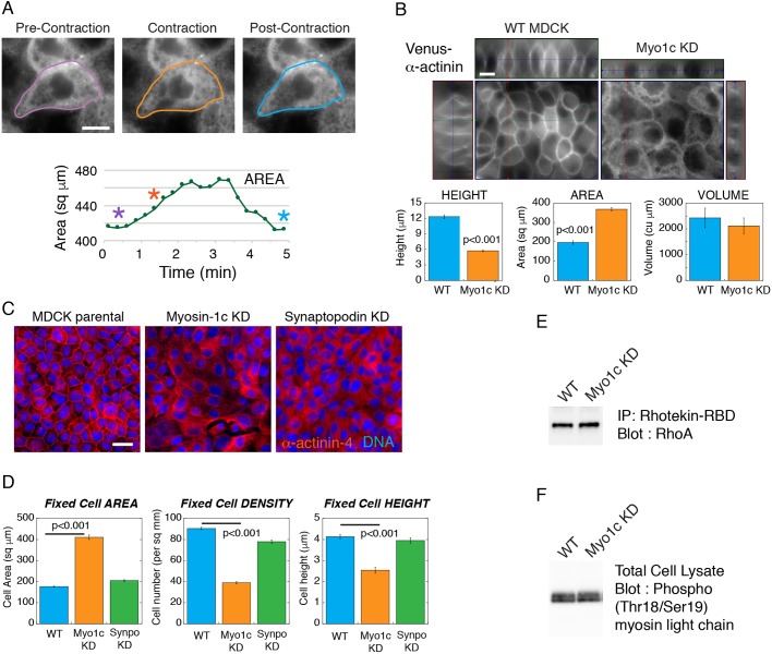
Myosin-1c knockdown compromises the coupling between actomyosin contractility and the lateral plasma membrane. (A) Live-cell images of Venus–α-actinin in the myosin-1c-knockdown (Myo1c KD) cell monolayer from Movie 7 showing pre-contraction (purple outline), contraction (orange outline) and post-contraction (blue outline) cell boundaries. The graph plots the area of the cell. Purple, orange and blue asterisks correspond to the cells outlined in purple, orange and blue, respectively. Data are representative of six sets of live-cell time-lapse movies. (B) Live-cell images of Venus–α-actinin in mature wild-type (WT MDCK) and myosin-1c-knockdown cell monolayers. x-z, y-z and x-y images were used to calculate the cell height, spread area and cell volume. Image set is representative of 16 sets of images from one experiment out of four independent experiments. Scale bars: 10 µm. Bar graphs show measurements of cell height, spread area and cell volume of wild-type and myosin-1c knockdown cells. Results are mean±s.e.m. of 16 measurements. P<0.001 for height and area between WT and myosin-1c KD. Data are representative of one experiment out of four independent experiments. (C) Wide-field immunofluorescence of α-actinin-4 in wild-type, myosin-1c knockdown and synaptopodin (Synpo) knockdown (KD) cell monolayers. Image set is representative of 16 sets of images from one experiment out of three independent experiments. Scale bar: 10 µm. (D) Cell spread area, cell density and cell height of wild-type, myosin-1c knockdown and synaptopodin knockdown monolayers. Bar graphs show the mean±s.e.m. of 16 measurements. P<0.001 for height, area and density between WT and myosin-1c KD. Data are representative of one experiment out of four independent experiments. (E) Immunoprecipitation of active Rho using the Rhotekin Rho-binding domain (RBD) followed by western blotting for Rho showing same level of Rho activities in MDCK wild-type and myosin-1c knockdown cells. Data are representative of one experiment out of three independent experiments. (F) Western blot for myosin light chain Thr18/Ser19 phosphorylation showing the same level in MDCK wild-type and myosin-1c knockdown cells. Data are representative of one experiment out of three independent experiments.

