Fig. 1.
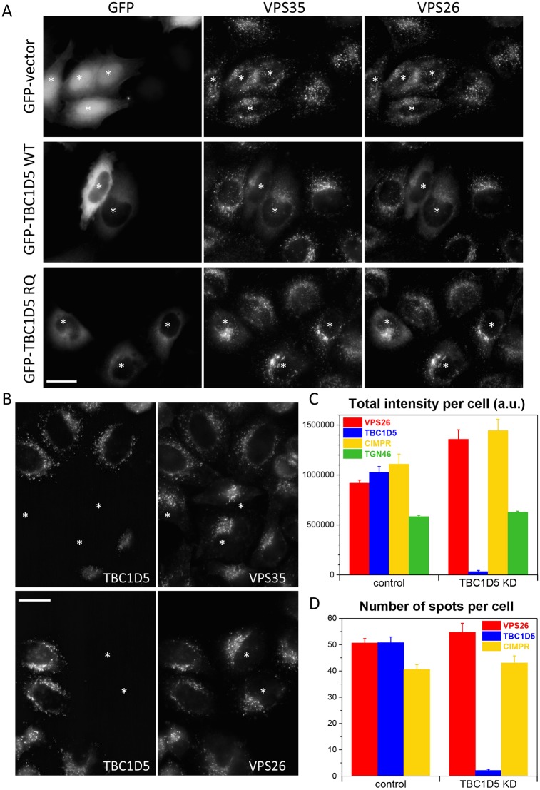
Loss of TBC1D5 expression enhances endosomal levels of the retromer CSC. (A) HeLa cells were transiently transfected with empty GFP vector, GFP-TBC1D5 wild type (WT) or GFP-TBC1D5 R169A/Q204A (RQ) mutant. After fixation, the cells were stained with antibodies against VPS35 and VPS26. Transfected cells are marked with an asterisk. Overexpression of the wild-type TBC1D5 can displace the retromer CSC from membranes. (B) HeLa cells were treated with siRNA to silence TBC1D5 expression. The knockdown cells were mixed with control cells and seeded onto coverslips. After fixation, cells were labelled with anti-TBC1D5 and antibodies against either VPS35 or VPS26. Loss of TBC1D5 expression (in cells marked with an asterisk) results in brighter staining of the retromer CSC proteins. (C,D) HeLa cells treated with siRNA to silence TBC1D5 expression were labelled with antibodies against VPS26, TBC1D5, CIMPR or TGN46 and then imaged using an automated microscope. Loss of TBC1D5 results in ∼40% increase in VPS26 fluorescence intensity but does not markedly increase the number of VPS26-positive spots (D). No spots were counted for TGN46 as the morphology of the TGN is not punctate but ribbon-like. P-values for TBC1D5 knockdown versus control: VPS26, 1.2×10⁻⁴; CIMPR, 0.0095; TGN46, 0.0037 for total intensity values. The P-values for spot numbers are: 0.08 for VPS26 and 0.23 for CIMPR. Scale bars: 20 µm.

