Fig. 7.

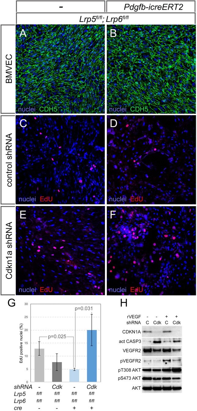
Wnt/β-catenin pathway interaction with CDKN1A and the apoptotic response. (A,B) BMVECs isolated from control (Lrp5fl/fl; Lrp6fl/fl) and conditional knockout (Pdgfb-icreERT2; Lrp5fl/fl; Lrp6fl/fl) mice labeled for nuclei (blue) and cadherin 5 (CDH5, green). (C-F) BMVECs from the indicated genotypes and shRNA treatments labeled for nuclei (blue) and EdU incorporation (red). (G) Quantification of EdU incorporation in BMVECs from the indicated genotypes and shRNA treatments (n≥3). Statistical significance was determined using two-way ANOVA with Bonferroni post-hoc test. Data are mean±s.e.m. (H) Immunoblots from lysates of BMVECs treated with combinations of recombinant VEGFA (rVEGFA) and an shRNA to Cdkn1a (C, control shRNA; Cdk, shRNA to Cdkn1a). In descending order, the immunoblots show detection of CDKN1A, activated caspase 3, VEGFR2, phospho-VEGFR2, phospho-T308-Akt, phospho-S473-Akt and total Akt.
