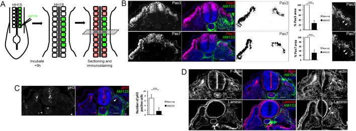
Fig. 2.
Dermomyotome growth, epithelial organization and basement membrane deposition are disrupted after inhibition of miR-133. (A) Schematic overview of the experimental approach. Posterior somites of HH14/15 embryos were injected with FITC-labelled antagomir-133 (AM133) and the downstream analysis was performed by immunostaining after 9 h of incubation. (B) Immunostaining for Pax3, Pax7, AM133 and DAPI as indicated. The areas positive for Pax3 or Pax7 within the somite were quantified using Fiji/ImageJ, and were significantly smaller in AM133-injected somites compared with somites from the noninjected contralateral control side. Higher magnification images of injected somites showed disruption to dermomyotome morphology (white arrowheads). (C) Immunostaining for pH3, AM133 and DAPI as indicated. The number of pH3-positive cells was significantly reduced in AM133-injected somites (white arrowheads) compared with somites from the contralateral side. ***P<0.001. (D) Immunostaining for F-actin, laminin, AM133 and DAPI as indicated. Higher magnification images of noninjected and injected somites stained for F-actin or laminin are shown on the right. White arrowheads indicate disorganized and disrupted staining in the dermomyotome region. Scale bars: 50 μm.

