. 2018 May 29;7:e34100. doi: 10.7554/eLife.34100

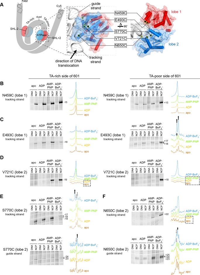
Figure 1. Interactions between the ATPase motor of Chd1 and nucleosomal DNA are sensitive to both nucleotide state and DNA sequence.

(A) Overview of the Chd1 ATPase motor on the nucleosome. A cartoon schematic (left) shows the superhelical wrap of nucleosomal DNA, with an ATPase motor (blue/red) at each of the superhelix location (SHL) 2 sites on either side of the nucleosome. The expanded views on the right show the molecular surface of the ATPase motor in complex with nucleosomal DNA, based on a cryoEM complex (Farnung et al., 2017). The five positions individually substituted with cysteine for APB cross-linking are highlighted with yellow circles, and the tracking and guide strands of DNA are indicated. The PyMOL visualization package (RRID:SCR_000305) was used for molecular rendering. (B-F). Representative data showing cross-linking to nucleosomal DNA on either the TA-rich side (left) or TA-poor side (right) of the 601 sequence, in different nucleotide conditions. Identical cross-linking reactions were performed with 300-450 nM Chd1 variant with either 150 nM canonical 601 nucleosomes with 40 bp of flanking DNA on each side ('NCP') or 150 nM naked DNA ('DNA'), to ensure observed cross-links were nucleosome-specific. Bands that increase or decrease in cross-linking efficiency for ADP·BeF3 compared to other conditions are indicated with black or white circles, respectively. Scans of each lane are shown as intensity plots to the right of each gel, with arrows highlighting bands that showed relative increases (black) or decreases (white) for ADP·BeF3 conditions. Dotted boxes highlight nucleotide conditions where cross-links were severely reduced or not detected. Numbering refers to the distances (nt) of cross-linked sites from the 601 dyad (positive numbers for TA-poor side and negative numbers for TA-rich side). These data are representative of 4 or more experiments. Extended gel images are shown in Figure 1—figure supplement 3.

Figure 1.

Figure 1—figure supplement 1. Site-specific cross-linking visualized on the Chd1-nucleosome structure.
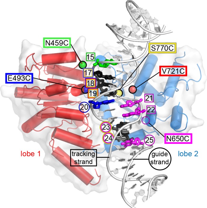
Figure 1—figure supplement 1.

This image shows the ATPase motor of Chd1 bound to nucleosomal DNA at SHL2 in the cryoEM structure 5O9G (Farnung et al., 2017), captured in an ADP·BeF3 state. Each residue used for site-specific cross-linking is highlighted with a colored sphere at the Cα position. Residue N650 was not built in the cryoEM structure (dotted magenta circle), and its position was therefore estimated based on superimposing the Chd1 crystal structure 3MWY (Hauk et al., 2010). DNA nucleotides are colored according to cross-links from each cysteine-substituted site. Nucleotides cross-linked by multiple sites are colored black, with colored circles (guide strand) or squares (tracking strand) indicating the origin of cross-linking. Cross-linking sites shown here represents a compilation of both TA-rich and TA-poor sides of the Widom 601 nucleosome. The PyMOL visualization package (RRID:SCR_000305) was used for molecular rendering.
Figure 1—figure supplement 2. DNA sequences of 601 variants used in this study.

Figure 1—figure supplement 2.

Sequence information for the core 145 bp of the canonical and variant 601 positioning sequences used in this study (5’ to 3’). Periodic TA steps are indicated with gray dotted boxes, and colored segments highlight sequence elements that are altered in 601 variants, corresponding to the cartoons in Figure 4. The red box indicates the inserted A/T bp for the variant 601[insert(+1) TA-rich]. These core segments were made into double-labeled Cy5/FAM 40N40 nucleosomes using primers that matched flanking DNA in the original Widom 601 plasmid: 5’-/Cy5/AGTTCATCCCTTATGTGATGGACCCTATAC on the TA-rich (left) side, and 5’-/6FAM/ATCCGACTGGCACCGGCAAGGTCGCTGTTC on the TA-poor (right) side.
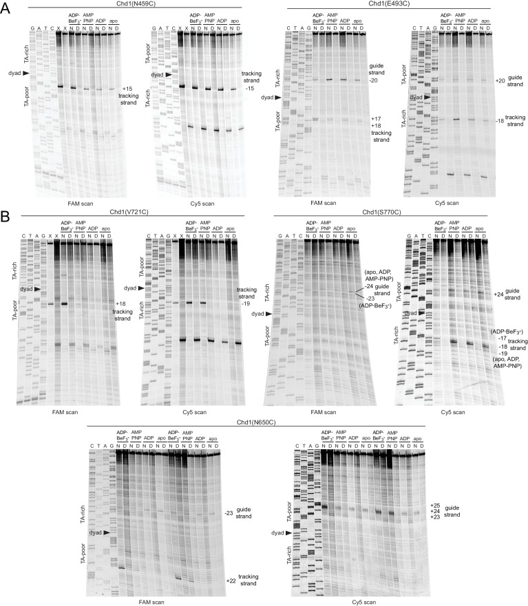
Figure 1—figure supplement 3. Extended gel images of Chd1 cross-linking reactions.

Figure 1—figure supplement 3.

Scans of urea denaturing gels, shown in Figure 1, for cross-linking reactions performed with cysteine substitutions in ATPase lobe 1 (A) or lobe 2 (B). Each reaction was carried out with either 40N40 nucleosomes (N) or naked DNA (D) with the canonical Widom 601 sequence. Numbering refers to the distance (nt) from the 601 dyad.
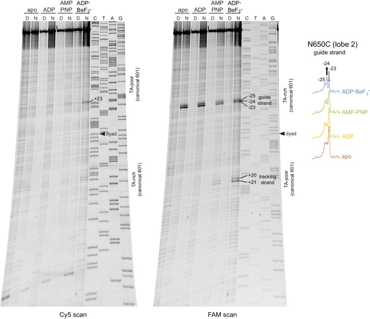
Figure 1—figure supplement 4. Chd1(N650C) cross-linking on 601[swap SHL2.5/3.5] nucleosomes yielded a distinct nucleotide-dependent pattern.

Figure 1—figure supplement 4.

Altering the Widom 601 sequence affects the cross-linking pattern of the Chd1 ATPase motor. The major cross-linked species for the 601[swap SHL2.5/3.5] nucleosome (−23 to −25 on the guide strand) were on the opposite side compared to the canonical 601 (+23 to +25 on the guide strand). This difference corresponds with the swap of 24–39 bp segments on either side of the dyad. In addition to switching sides, cross-linking to the 601[swap SHL2.5/3.5] also displayed a different nucleotide- dependent pattern. For this nucleosome, cross-linking in ADP·BeF3 was enhanced at position −24 and diminished one nt closer to the dyad (−23) compared to the other nucleotide states. This contrasts with the canonical 601, which also showed stronger cross-linking in ADP·BeF3 at +24 compared to other nucleotide states, but had decreased cross-linking farther from the dyad at +25 (Figure 1).