. 2018 May 29;7:e34100. doi: 10.7554/eLife.34100

Figure 3. The Chd1-dependent shift in histone-DNA cross-linking correlates with Chd1 binding to the adjacent SHL2, on the same DNA gyre of the nucleosome.

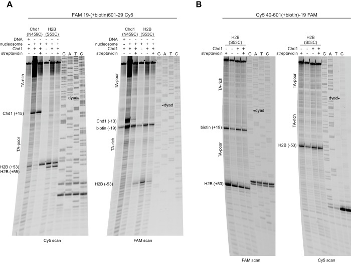
(A) The presence of a biotin/streptavidin block at the TA-rich SHL–2 site does not diminish the Chd1-dependent shift in H2B(S53C) cross-links on the TA-poor side. The nucleosome substrate for these experiments was 19-[+biotin]601-29, with a biotin moiety 19 nt from the dyad on the TA-rich SHL–2 site. Note that for this nucleosome, the FAM and Cy5 labels are on opposite sides compared to all other 601 nucleosomes used in this study. Chd1(N459C) cross-linking experiments monitored Chd1 binding at each SHL2, and H2B(S53C) experiments followed the Chd1-dependent shift in histone-DNA cross-links on the TA-poor side. The apparent shift in Chd1(N459C) cross-linking site on the biotinylated strand (labeled as −13) was due in part to slower migration caused by the biotin moiety on the DNA fragment, and may not represent a true change in cross-linking position. The gels are representative of three or more experiments, and extended gel images are shown in Figure 3—figure supplement 1A. (B) The presence of a biotin moiety at the TA-poor SHL+2 site prevents the Chd1-dependent shift in H2B(S53C) cross-linking on the TA-poor side. The nucleosome substrate for these experiments was 40-601[+biotin]-19, with a biotin moiety 19 nt from the dyad on the TA-poor SHL+2 site. The absence of the H2B(S53C) shift likely stems from poor binding due to the biotin, as shown in Figure 3—figure supplement 2. The gels are representative of three or more experiments, and extended gel images are shown in Figure 3—figure supplement 1B. (C) A model for how Chd1 binding at SHL+2 may be coupled to shifts in DNA cross-linking.

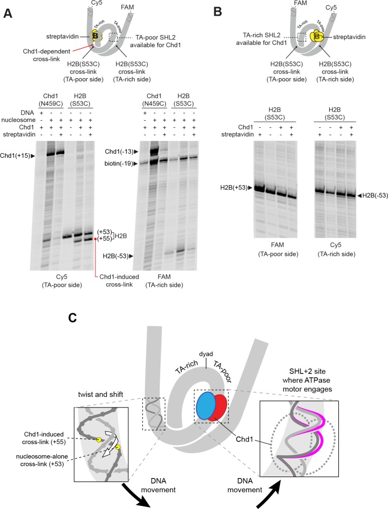
Figure 3.

Figure 3—figure supplement 1. Extended gel images for H2B(S53C) cross-linking experiments with biotinylated 601 nucleosomes.

Figure 3—figure supplement 1.

Scans of urea denaturing gels, shown in Figure 3, for reactions using nucleosomes that contained a biotin moiety 19 nt from the dyad. (A) Experiment using 19-(+biotin)601–29, which was biotinylated on the TA-rich side, as shown in Figure 3A. (B) Experiment using 40-601(+biotin)−19, which was biotinylated on the TA-poor side, as shown in Figure 3B. These experiments are representative examples of three or more independent experiments.
Figure 3—figure supplement 2. A biotin moiety on the TA-poor SHL2 interferes with Chd1 binding, independently of streptavidin.

Figure 3—figure supplement 2.

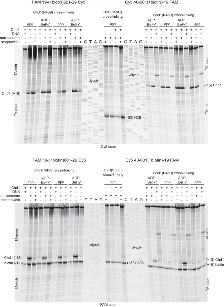
Shown are duplicate experiments for Chd1(N459C) cross-linking, using nucleosomes with a biotin on the TA-rich SHL–2 (19-[+biotin]601–29, left) or the TA-poor SHL+2 (40-601[+biotin]−19, right). For both of these nucleosomes, biotin is 19 nt from the dyad on the tracking strand, and the numbers of the construct name indicate the lengths of DNA flanking the core 601 sequence. Note that the FAM label is on the same side as the biotin, so the TA-rich and TA-poor sides appear in opposite orientations for these two nucleosomes for each scan. The Cy5 scan (top) shows that in the absence of a biotin moiety, Chd1(N459C) cross-links strongly at the TA-rich and TA-poor SHL2 sites. In contrast, the FAM scan (bottom) shows a marked decrease in Chd1(N459C) cross-linking with biotin on the TA-poor side in apo but not ADP·BeF3 conditions. The middle lanes show a replicate experiment monitoring H2B(S53C) cross-linking for the 40-601[+biotin]−19 construct, where the biotin at the TA-poor SHL2 prevents a Chd1-induced shift.
Figure 3—figure supplement 3. Mutation of a conserved tryptophan (W793A) in the ATPase core of Chd1 only modestly slows the rate of nucleosome sliding and does not interfere with the two nt shift of H2B(S53C).

Figure 3—figure supplement 3.

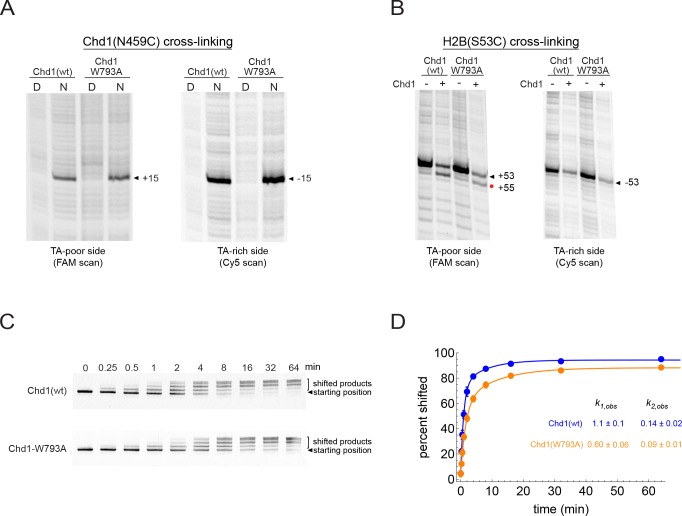
(A) The W793A mutation does not interfere with ATPase binding at SHL2. Shown are cross-linking reactions for Chd1(N459C) with and without the W793A mutation. Cross-linking was performed without nucleotide. No differences in cross-linking are apparent. (B) The W793A mutation does not disrupt the Chd1-dependent shift in H2B(S53C) cross-linking on the TA-poor side. These cross-linking reactions were carried out with the double mutant variant Chd1(N495C/W793A) under apo conditions. (C) Native gel sliding assay, showing repositioning of nucleosomes over time by wildtype Chd1 and a Chd1(W793A) variant. In this experiment, 50 nM Chd1 was incubated with 150 nM 0N80 nucleosome in the presence of 2.5 mM ATP and 5 mM MgCl2. (D) Quantitation and fitting of native gel sliding data shown in C. Colored circles are the averages of five replicates, with standard deviations shown as error bars (often obscured markers). Data were fit to a double exponential, and for the faster rate, Chd1(W793A) was approximately 2-fold slower than wild type Chd1.