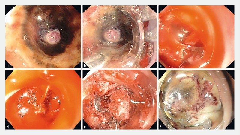
Fig. 2.

Cause and solution of incomplete hemostasis after OTSC placement. a nonsteroidal anti-inflammatory drug-related duodenal ulcer with a large visible vessel was seen in the distal duodenal bulb of an elderly patient with metastatic lung cancer. The ulcer was located within a narrowed lumen. b A therapeutic endoscope with 6-mm channel that was equipped with a large OTSC was used (OD of OTSC = 21 mm). The tip of the OTSC anchor was placed next to the visible vessel. c After opening the anchor, the ulcer was pulled and simultaneously suctioned into the OTSC, and the clip was released. d A large stream of blood was seen flowing from the artery. The clip was misplaced. The endoscope was left to suction the blood to prevent formation of large clots and to maintain visualization, while another therapeutic endoscope was being equipped with a medium-size OTSC. e The bleeding vessel was suctioned into the OTSC. The clip was placed ideally. Bleeding ceased instantly. f There was no further bleeding. The clip at 24 hours.
