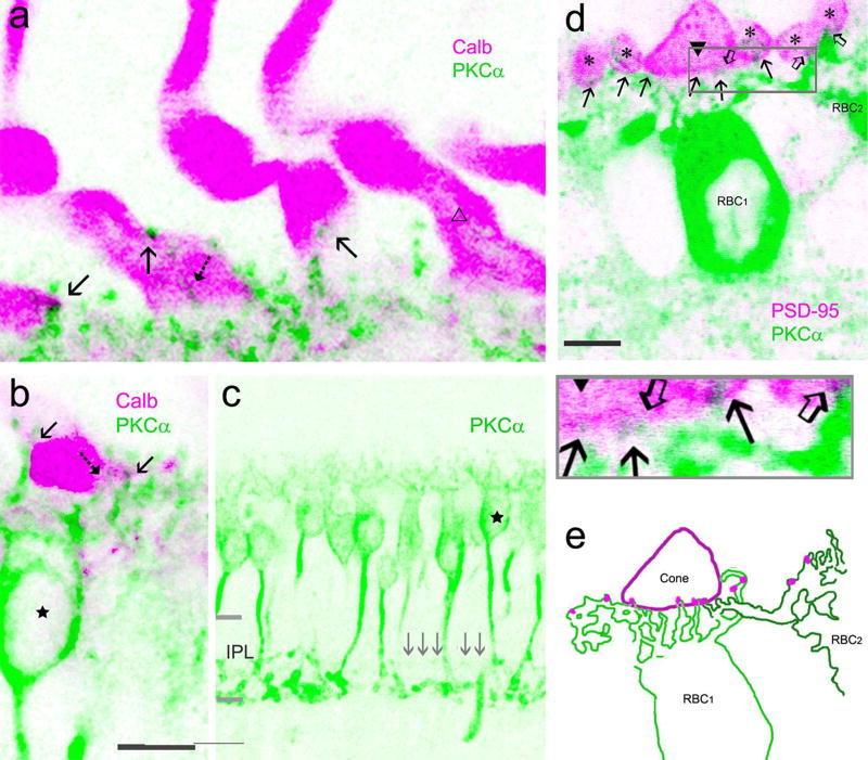
Fig. 7.
Confocal microscopy of baboon cone-RBC contacts. In a to b, cones are positively labeled by calbindin D-28k (Calb, pink). Each cone shows a tall fat sock-like pedicle with two vertically arranged segments. PKCα-labeled RBC dendrites (green) contact the lower segment, mostly by superficial contacts (arrow) and sometimes by invaginating contact (dashed arrow). Some cone pedicles do not contact RBCs (open triangle, a). The axon of the RBC (star, b and c), like those of other RBCs, branches at ~50–70% of the IPL depth and terminates near 100% of the IPL depth. The signals (gray arrows) restricted to the center of the IPL, presumably axons of DB4 cone bipolar cells, are almost invisible. In d, PKCα-labeled dendrites (green) contact both a cone pedicle (closed triangle) and rod spherules (asterisks) labeled by PSD-95 (pink), and these contacts (arrows for RBC1, open arrows for RBC2) are manually traced from the image and displayed in e (pink dots). IPL-the inner plexiform layer. The scale bar is 5 µm in a and d and 10 µm in b.

