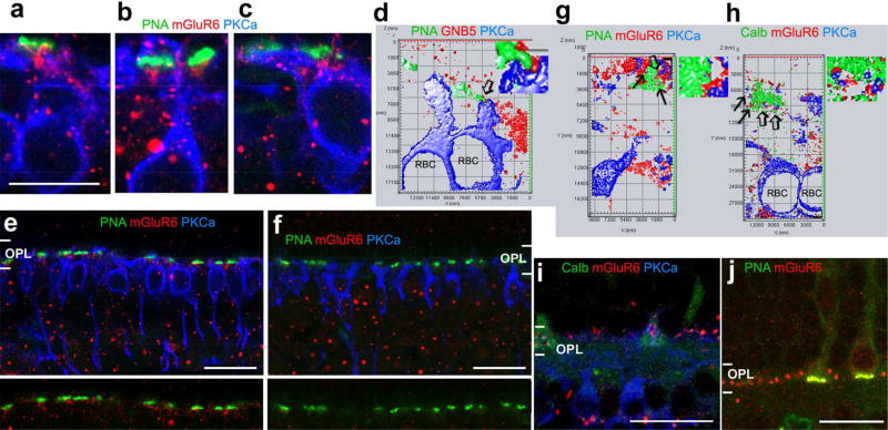
Fig. 8.
GNB5 and mGluR6 expressed in the contacts between cones and RBCs. Mouse (a–f) and baboon (g–j) retinal slices were double- or tripled-labeled. Synaptic contacts were studied in 1–2-µm-thick blocks with a resolution of 30 nm per pixel (d, g and h) and in series of optical sections ≤1 µm-thick (a–c, e and f). d, g and h display the 3D surface profile reconstructed from a series of optical sections with a step of 150–180 nm. GNB5 and mGluR6 (red) are present in contacts (arrow) formed by PKCα-labeled RBC dendrites (blue) and PNA or calbindin (Calb)-labeled cone pedicles (green) (a–d, g and h). Goat anti-mGluR6 (g, h and i) and rabbit anti-mGluR6 (the rest images) label primarily the outer plexiform layer (OPL) in the monkey retina (g to j). In the mouse retina, they label the OPL and inner nuclear layer (e), but the immunoreactivity in the OPL is absent in the mGluR6 knockout mouse (f). The distribution of GNB5 immunoreactivity in the OPL is like mGluR6 immunoreactivity (d). Small regions pointed by open arrows in d, g and h are amplified and depicted to the right. GNB5-G protein beta 5. The scale bar is 10 µm for a–c and 20 µm for e, f, i and j.

