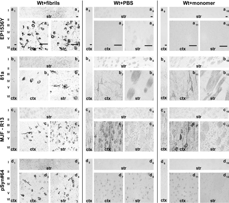
Fig. 3. Assessment of pSer129-α-Syn and inclusion detection in rat brain.
Wild-type Sprague Dawley rats (SDTac) were injected bilaterally into the striatum with 20 µg of α-syn pre-formed fibrils or monomeric protein as previously described(Abdelmotilib et al., 2017). Brain sections were evaluated from rats six months later. Representative widefield images of DAB-staining (converted to grayscale for contrast) with the indicated monoclonal antibody (left margins) in animals injected with A) fibrils in wild-type rats, B) saline control in wild-type rats, and C) monomer protein in wild-type rats. All antibodies were included at 10 ng mL−1 concentration. Coronal sections are shown within ~1 mm of Bregma, with primary motor cortex (ctx) and dorsal striatum (str) shown at two different magnifications. Scale bars are 0.1 mm.

