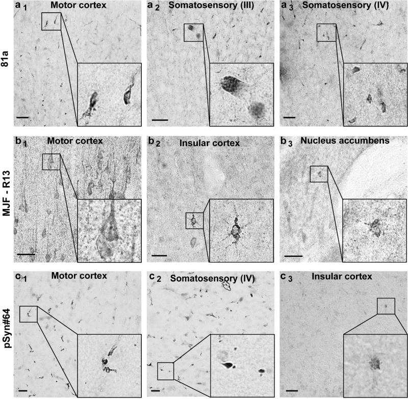
Fig. 4. Assessment of pSer129-α-syn monoclonal antibodies in α-syn knockout mice.
Coronal brain sections were obtained from adult (3–6 months) α-syn knockout mice and evaluated with the monoclonal antibody indicated in the left margin (a, b, c), all used at 10 ng mL−1. Representative images from DAB staining (grayscale shown for contrast) are shown. Insets black bounding boxes show higher magnifications of features of interest. Scale bars are 0.1 mm.

