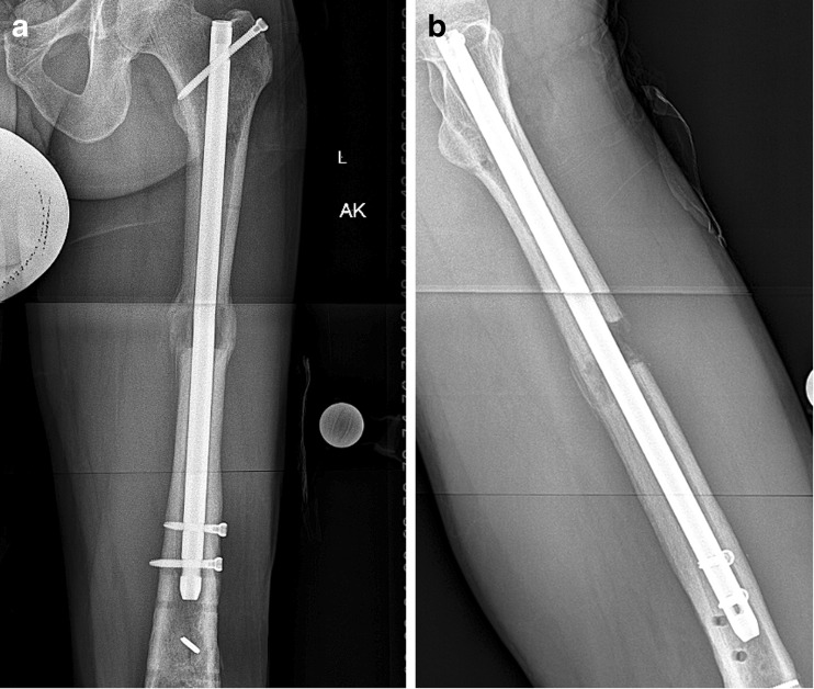
Fig. 6.
a This AP radiograph of the same case as Figs. 2, 3, and 4, post-distal nail locking and frame removal, shows two cortices of bridging callus. b The lateral X-ray of the same patient shows bridging callus posteriorly but not anteriorly. With bony bridging on the posterior, medial, and lateral cortices, this patient is considered united.

