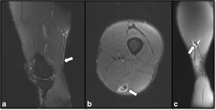
Fig. 1.
Preoperative magnetic resonance imaging (MRI) depicting the distal semitendinosus injury. Sagittal T2, fat suppressed MRI image showing proximal retraction of the distal semitendinosus tendon (arrow) (a). Axial T2, fat suppressed MRI image showing injury to the semitendinosus with surrounding scar formation (arrow) (b). Coronal T2, fat suppressed MRI depicting the injury at the musculotendinous junction of the semitendinosus (arrow) (c).

