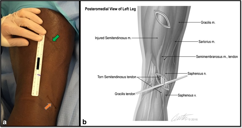
Fig. 2.
A two-incision surgical technique is utilized for isolating the injured semitendinosus. Using preoperative measurements with magnetic resonance imaging, surgical incisions are marked at the level of the retracted distal tendon (orange arrow) and proximal musculotendinous junction (green arrow) of the semitendinosus over the posteromedial thigh (a). An artistic rendering of the surgical approach, with relevant anatomy encountered through both incisions (b).

