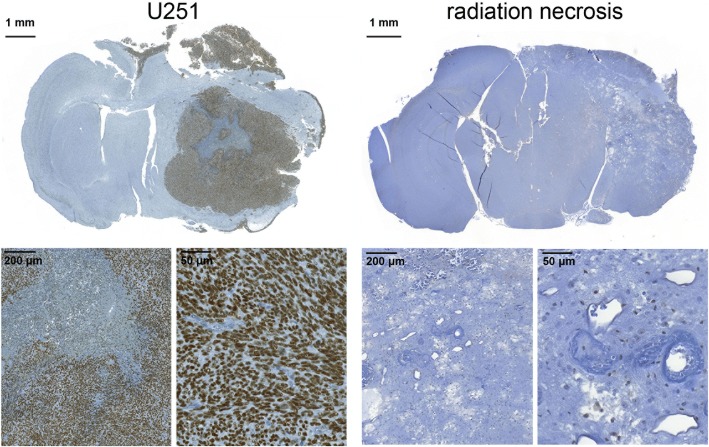
Fig. 2.
Anti-PARP1 immunohistochemistry. Staining of transaxial formalin-fixed, paraffin-embedded sections of mice implanted with U251 tumors (left group) mice with experimental radiation necrosis (right group) reveals high PARP1 expression in the nuclei of tumor cells and low PARP1 expression elsewhere in healthy brain and radiation necrosis

