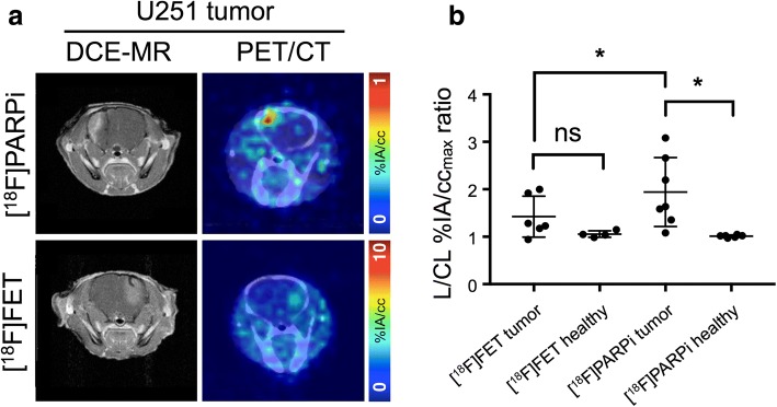
Fig. 5.
PET imaging of mice with focal intracranial U251 cell xenografts. a (left column) DCE-MR and (right column) fused PET/CT transaxial slices of mice with U251 tumor, injected with (top row) [18F]PARPi and (bottom row) [18F]FET. b Lesioned-to-contralateral hemisphere %IA/ccmax ratios for mice in different groups. *Significant at p < 0.05

