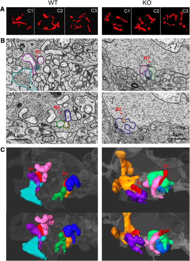
Figure 5.

Aberrant synaptic organization of cone synapses in α2δ-4 KO mice determined by serial block face scanning EM. A, Examples of ribbons traced from sections through 3 cone pedicles (c1–c3) from WT and α2δ-4 KO retina. B, Two image planes (top, bottom) of a WT (left) or KO (right) cone pedicle each with 2 ribbons (R1, R2). Processes that contact the cone at these ribbon sites are pseudocolored. Each process was traced to some distance from the ribbon site to provide a 3D view of the synaptic arrangement at the ribbon site. C, Two 3D views (top, bottom) of the processes colored in B and their associated ribbon (R1, R2). Left, In the WT pedicle, typical triad-like arrangements are observed at both ribbon (R1, R2) sites, as evidenced by three processes converging at the ribbon site. The identities of these processes are not confirmed because of their partial reconstructions. Similar results were obtained in reconstructions of HET cone pedicles (Fig. 5-1). Right, In the KO pedicle, triadic arrangement of processes at the ribbon sites was not readily apparent. The R1 ribbon terminated opposite two processes (magenta, green), with a third (blue) somewhat further away. The R2 ribbon was apposed to only two processes (orange, purple), and did not appear “sandwiched” by these two processes, as typically seen in WT triads.
