
Figure 5.
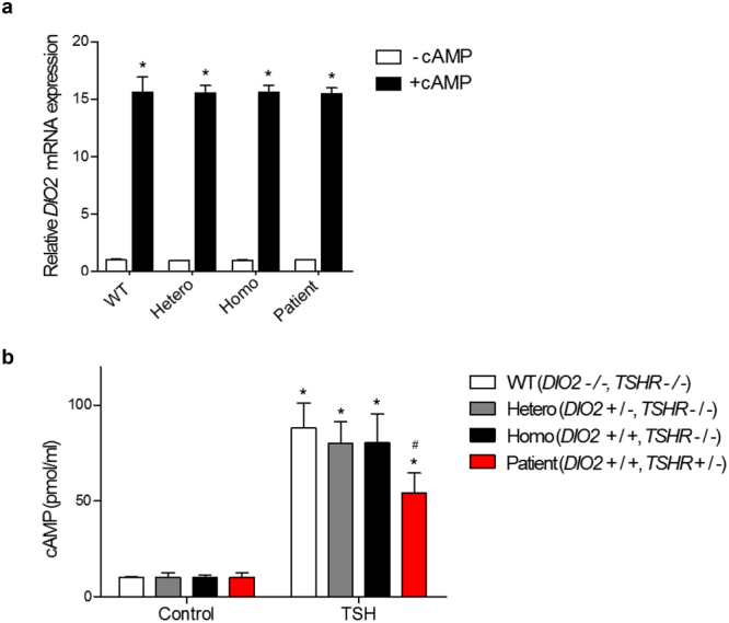
Relative DIO2 mRNA expression levels in cAMP-treated cells and cAMP assay results in TSH-treated in primary-cultured skin fibroblast cells. (a) Cells are treated with 1 mM db-cAMP for 16 h (+cAMP) and total RNA is extracted from the cells and DIO2 mRNA expression level is analyzed by quantitative real-time RT-PCR. The cAMP-non-treated cells are used as negative control (−cAMP). DIO2 mRNA expressions in WT, Hetero, Homo and patient are significantly increased by cAMP treatment. *p < 0.05 vs. negative control in each tested fibroblast. (b) Cells were treated with 1 IU of TSH for 30 min and then are treated with 0.1 N hydrochloric acid to stop endogenous phosphodiesterase activity. After cell lysis, intracellular cAMP concentration is measured using cAMP direct immunoassay kit. The TSH-non-treated cells are used as negative control. cAMP is increased in all groups, but significantly less increased in the patient. *p < 0.05 vs. negative control in each tested fibroblast. #p < 0.05 vs. WT.