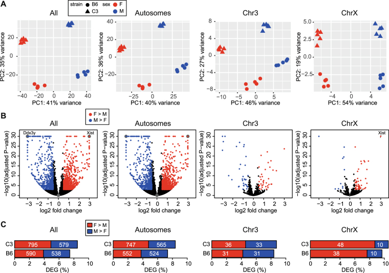
Figure 1.
Mechanisms of dosage compensation maintain female to male expression balance for the majority of genes on the X chromosome. (A) Principal component analysis (PCA) of gene expression using RNA-seq data from all chromosomes, autosomes, chromosome 3, and chromosome X. Each point represents a liver sample (n = 5 mice per group) with color indicating sex and shape indicating strain. Percent of variance for each principal component (PC1, PC2) is indicated on the axes. (B) Volcano plots depicting sex-specific differential expression of genes on all chromosomes, autosomes, chromosome 3, and chromosome X for C3 mice. Significant genes (adjusted P-value < 0.001; |log2 fold change >0.5|) are colored by direction of fold change (Red, female-bias; Blue, male-bias). For visualization purposes, adjusted P-values less than 1.0e-30 were set to 1.0e-30, log2 fold change <−3 were set to −3, and log2 fold change >3 were set to 3. Overlapping points are visualized using sunflower petals. Female-specific Xist (chromosome X) and male-specific Ddx3y (chromosome Y) are labeled. (C) Barplots depicting the percent of female- (red) and male-biased (blue) differentially expressed genes (DEGs) on all chromosomes, autosomes, chromosome 3, and chromosome X. The absolute number of DEGs is indicated on each bar. ChrX, chromosome X; Chr3, chromosome 3.

