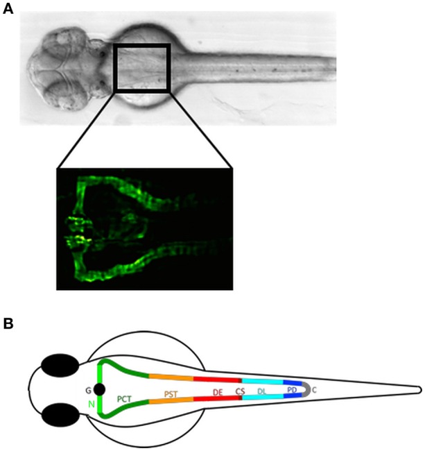
Figure 1.

The zebrafish pronephros: anatomical position and segmental organization. (A) Brightfield dorsal view of a 2 day post fertilization (dpf) zebrafish embryo (upper panel). The rectangle in the anterior trunk indicates the location of the proximal pronephric structures with a fused glomerulus at the midline that connects to the segmented pronephric tubules as labeled in the Tg(wt1b:egfp) zebrafish line by GFP expression (lower panel). (B) Schematic illustration of a zebrafish pronephros showing segmental organization of each nephron into glomerulus (G), neck (N), proximal convoluted tubule (PCT), proximal straight tubule (PST), distal early (DE), corpuscle of Stannius (CS), distal late (DL), and pronephric duct (PD) that fuse to the cloaca (C). Adapted from Wingert and Davidson (34).
