
Figure 3.
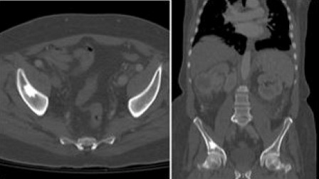
CT Scan Demonstrating Patient with Low Volume Bony Disease. Low volume disease: CT scan with I.V contrast axial and sagittal bone window. Single sclerotic bone lesion involving right iliac bone.
Official websites use .gov
A
.gov website belongs to an official
government organization in the United States.
Secure .gov websites use HTTPS
A lock (
) or https:// means you've safely
connected to the .gov website. Share sensitive
information only on official, secure websites.
CT Scan Demonstrating Patient with Low Volume Bony Disease. Low volume disease: CT scan with I.V contrast axial and sagittal bone window. Single sclerotic bone lesion involving right iliac bone.