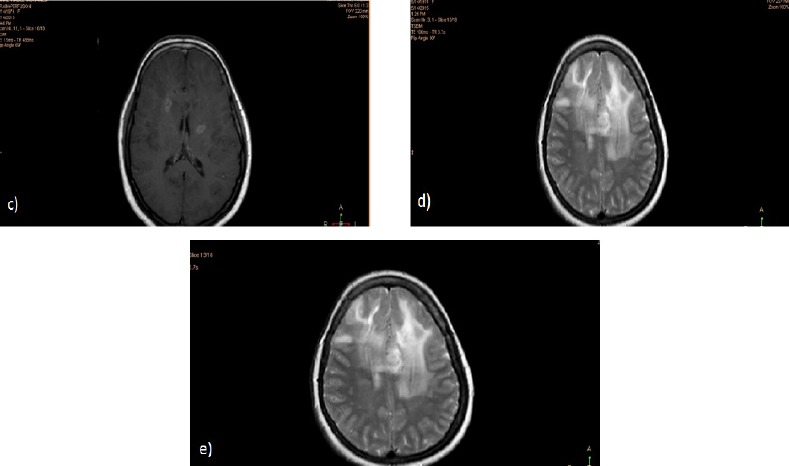
Figure 1c, d and e.

Post Contrast Axial T1 (c and d) and FLAIR (e) Weighted Images Showing Evidence of Newly Developed Well-Defined Lobulated Lesion at the Left High Parietal Region, Periventricular in Location, Involving the Corpus Callosum, Appearing of High T2 Signal Intensity with Heterogeneous Post Contrast Enhancement. It is seen surrounded by high T2 and FLAIR signal suggestive of vasogenic edema. Similar but smaller lesion is also noted at the right high parietal region
