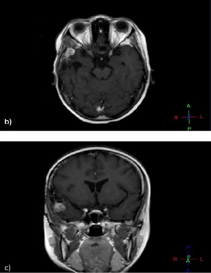
Figure 2b and c.

On Her Regular Follow up, Post Contrast Axial and Coronal T1 Weighted Images Showing Newly Developed Right Temporal Enhancing SOL at the Operative Bed.

On Her Regular Follow up, Post Contrast Axial and Coronal T1 Weighted Images Showing Newly Developed Right Temporal Enhancing SOL at the Operative Bed.