
Figure 2.
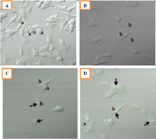
Morphology of MCF-7 Cells after 8 Weeks of Treatments. Cells were seeded at density of 104 viable cells/ml per well. Observations were done using confocal fluorescent microscope at 40x magnification. A: control (DMSO), B: End+BE, C: End+BE+Cur 8.5, D: End+BE+Cur17. Green fluorescence indicated the location of curcumin in a cell (black arrow); a: cobblestone appearance; b: fibroblast-like appearance