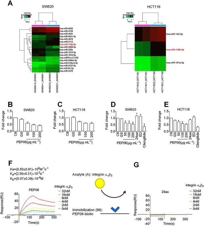
Figure 4.

PEP06 down‐regulated the expression of miR‐146b‐5p by forming high‐affinity interactions with αvβ3 integrin in CRC cells. (A) Heat map of microRNA microarray analysis showing the alterations in the expression of miRNAs in SW620 and HCT116 cells exposed to 200 μg·mL−1 PEP06 for 24 h relative to control cells. SW620C1, SW620C2, HCT116C1 and HCT116C2 represent control samples in duplicate, and SW620 + 1, SW620 + 2, HCT116 + 1 and HCT116 + 2 represent PEP06‐treated samples in duplicate. (B, C) qRT‐PCR verification of miR‐146b‐5p down‐regulation in SW620 and HCT116 cells treated with varying concentrations of PEP06. Data (mean ± SEM) were from eight independent experiments. * P < 0.05 by one‐way ANOVA, Dunnett's test: compared with Ctl. (D, E) qRT‐PCR verification of miR‐146b‐5p down‐regulation in SW620 and HCT116 cells treated with PEP06 compared with 24aa, Endostar and cilengitide. Data (mean ± SEM) were from five independent experiments. * P < 0.05 by one‐way ANOVA, Dunnett's test: compared with Ctl. (F) Sensorgrams of αvβ3 integrin (1–32 nM) binding to immobilized PEP06 (50 nM) and K D values calculated. The kinetic parameters were measured by Biacore evaluation software. (G) SPR sensorgrams showing no binding of αvβ3 integrin (1–32 nM) to immobilized 24aa (50 nM).
