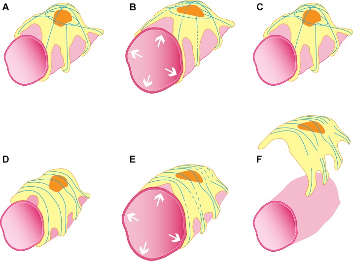
Fig. 1.
Schematic illustration hypothesizing the in vivo interaction between glomerular capillary expansion-induced stretch in wild-type (A–C) and FSGS-causing point-mutant ACTN4 podocytes (E–F). In response to a transient stretch, wild-type podocytes develop small areas of actin breakages (represented by blue lines; see B) that repair (C), allowing the podocyte to maintain its contraction and remain adhered to the glomerular basement membrane. In contrast, mutant ACTN4 podocytes show breakages in their actin structure after stretch (E) that fail to repair (F). Hence, the podocyte’s contraction fails in association with its detachment.

