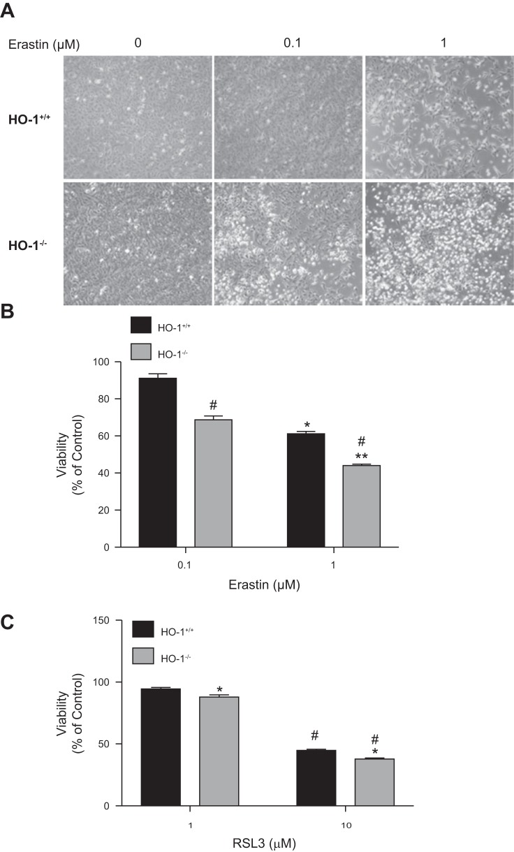
Fig. 3.
Lack of HO-1 increases PTC sensitivity to erastin-induced ferroptosis. A: phase contrast microscopy images of HO-1+/+and HO-1−/− PTCs treated with either 0.1 or 1 μM erastin for 16 h. B: cell viability after treatment of PTCs with 0.1 or 1 μM erastin or 1; *P < 0.05 compared with 0.1 μM erastin-treated HO-1+/+ PTCs; **P < 0.01 compared with 0.1 μM erastin-treated HO-1−/− PTCs; #P < 0.01 compared with erastin-treated HO-1+/+ PTCs. C: cell viability after treatment of PTCs with 10 μM RSL3 for 16 h; *P < 0.01 compared with 1 μM RSL3 treatment; #P < 0.05 compared with RSL3-treated HO-1+/+ PTCs. Data shown represent means ± SE of three independent experiments with four to six replicates in each experiment.

