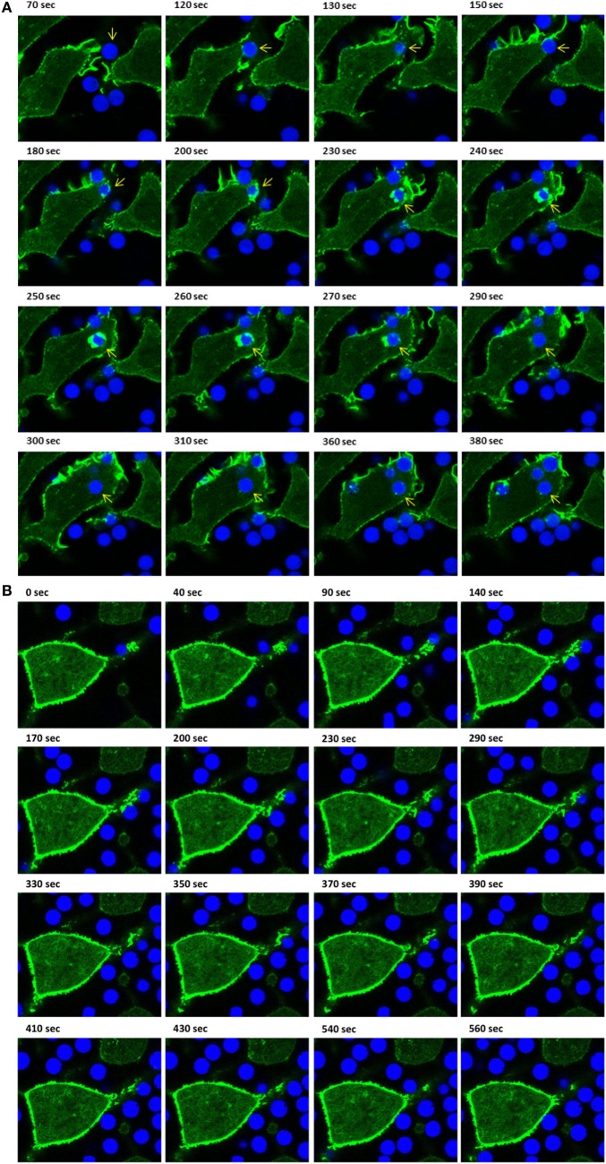
Figure 3.
Live-cell imaging of IpLITR 2.6b/IpFcRγ-L-mediated phagocytosis at different incubation temperatures. Rat basophilic leukemia-2H3 cells (3 × 10⁵) stably co-expressing IpLITR 2.6b/IpFcRγ-L and LifeAct-GFP were incubated at 37°C (A) or at 27°C (B) with 9 × 10⁵ αHA monoclonal antibody-coated 4.5 µm bright blue microspheres. Immediately after the addition of target beads, images were collected at 10 s intervals for ~8 min using a Zeiss LSM 710 laser scanning confocal microscope (objective 60×, 1.3 oil plan-Apochromat; Munich, Germany). Representative time-stamps in (A) were extracted from Video S3 in Presentation 2 of Supplementary Material and the time-stamps in (B) were from Video S4 in Presentation 2 of Supplementary Material. In (A), the target microsphere of interest is indicated with an arrowhead.

