Fig. 2.
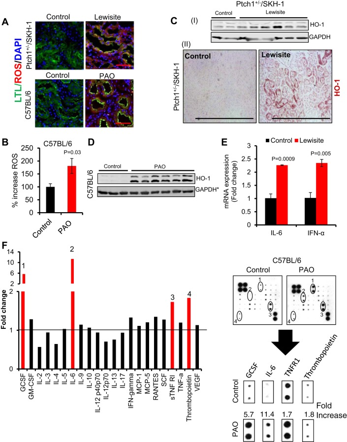
Cutaneous exposure of lewisite and PAO induces oxidative stress and inflammatory cytokines in the kidney. A: fluorescence-based photomicrograph analysis of reactive oxygen species (ROS) generation in the proximal tubules of kidneys of lewisite-treated Ptch1+/−/SKH-1 or PAO-treated C57BL/6 mice as assessed using CellROX Deep Red Reagent (red intensity). Proximal tubules were stained using fluorescence-conjugated Lotus tetragonolobus lectin (LTL) (green intensity). Bars = 25 µm. B: histogram showing plate reader-based quantification of ROS generation in the kidney lysates of vehicle- and PAO-treated C57BL/6 mice. n = 5. C: representative Western blot analysis of heme oxygenase-1 (HO-1) in kidney tissue lysates (CI) and IHC staining (CII). Bars = 50 µm in kidney tissue sections of vehicle- and lewisite-treated Ptch1+/−/SKH-1 mice. D: representative Western blot analysis of HO-1 in kidney tissue lysates of vehicle- and PAO-treated C57BL/6 mice. The lower band is ~28 kDa. *GAPDH loading control represent stripping of same blot as shown in Fig. 1G. E: RT-PCR analysis of mRNA expression of IL-6 and interferon (IFN)-α in kidney tissue of vehicle control and lewisite-exposed Ptch1+/−/SKH-1 mice. F: mouse cytokine antibody array expression patterns of different cytokines in serum samples of vehicle control and PAO-exposed C57BL/6 animals. Cytokines showing >1.5-fold induction [granulocyte colony-stimulating factor (GCSF) (1), IL-6 (2), tumor nerosis factor-α receptor (TNFR) 1 (3), and thrombopoietin (4)] are presented separately from the array images. In this experiment, serum samples of 3 mice from each group (control vs. PAO) were pooled and incubated with mouse cytokine antibody array membrane. Densitometry data obtained from the array images were analyzed by Excel-based analysis software tools (Ray Biotech). GM-CSF, granulocyte macrophage colony-stimulating factor; MCP, monocyte chemoattractant protein; RANTES, regulated on activation normal T cell expressed and secreted; SCF, stem cell factor; STNF RI, soluble tumor necrosis factor receptor I; VEGF, vascular endothelial growth factor.

