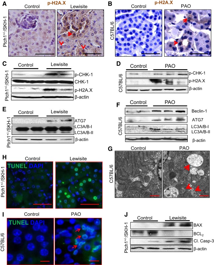
Fig. 3.
Cutaneous exposure to lewisite or PAO induces autophagy, DNA damage, and apoptosis responses in the kidney. A and B: immunohistochemical analysis showing enhanced nuclear staining of phosphorylated (p)-H2A.X (red arrows) in the tubules of the kidney tissue of lewisite-treated Ptch1+/−/SKH-1 mice or PAO-treated C57BL/6 mice compared with vehicle-treated controls. Bars = 25 µm. C and D: representative Western blot analysis shows enhanced phosphorylation-dependent activation of checkpoint kinase (CHK) 1 and H2A.X in the kidney tissue of lewisite (C)- and PAO (D)-treated mice compared with their respective control tissues. E and F: representative Western blot analysis of autophagy-related gene (ATG) 7 and LC3A/B-II in the kidney tissue lysates of lewisite-treated Ptch1+/−/SKH-1 mice (E) and beclin-1, ATG7, and LC3A/B-II in PAO-treated C57BL/6 mice (F) compared with their respective controls. G: transmission electron microscopy (TEM) analysis of kidney tissues of vehicle- and PAO-treated C57BL/6 mice (n = 3/group). Bars = 150 nm. Arrows indicate autophagosomes with damaged organelles and mitochondrial cristae loss. H and I: terminal deoxyribonucleotide-transferase-mediated dUTP nick-end labeling (TUNEL) staining of the kidney sections showing frequent presence of green TUNEL-positive cells in lewisite-treated (H, bars = 25 µm) and PAO-treated (I, bars = 12.5 µm) animals compared with vehicle-treated control animals. Apoptotic cells (green positive nuclei) were present in the tubules of the kidney. J: representative Western blot analysis showing induction of proapoptotic proteins, BAX and cleaved caspase-3, and reduced expression of anti-apoptotic BCL2 in the kidneys of lewisite-treated mice. β-Actin was used as a loading control. Western blot data shown in C, E, and J represent pooled kidney lysates for each group (n = 5, control vs. lewisite).

