Fig. 3.
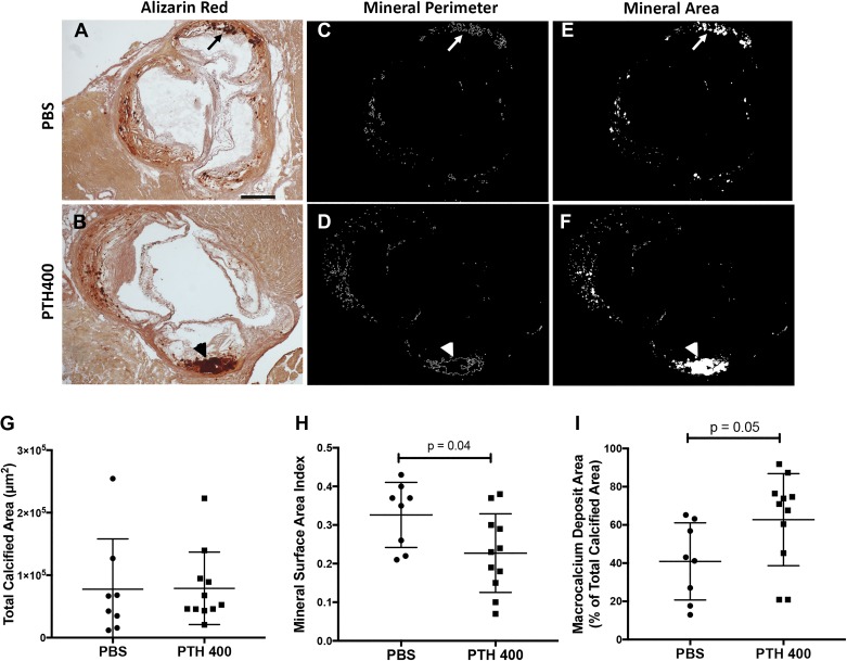
Effects of teriparatide on morphology of aortic calcium deposits. A and B: representative Alizarin red-stained aortic root sections from PBS-treated mice (A) and 400 µg/kg teriparatide (PTH400)-treated mice (B). The arrow in A denotes a region containing microcalcium deposits, whereas the arrowhead in B denotes a macrocalcium deposit. Scale bar = 500 µm. C–F: calcified deposits were computationally segmented, and their total perimeters (C and D) and areas (E and F) were drawn and measured in their respective groups. G–I: comparison of the total calcified area (G), mineral surface area index (total perimeter/total area; H), and percentage of calcified area present in macrocalcium deposits between groups (I). Statistical analysis was performed using a two-tailed Student’s t-test.

