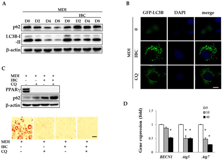
Figure 5.
Effect of IBC on autophagic flux during adipocyte differentiation. (A) 3T3-L1 adipocytes were differentiated in the presence or absence of 40 μM of IBC. Differentiating adipocytes were harvested at the indicated period and lysed for Western blotting analysis to determine levels of LC3B and SQSTM1/p62; (B) Preadipocytes were transfected with GFP-LC3 plasmid and differentiated with MDI in the presence of IBC or CQ (10 μM) during D0–D2, followed by additional treatment with differentiation medium for 2 days (D4). Cells were fixed and stained with DAPI (nuclei, blue-colored). Intracellular GFP-LC3 puncta (green-colored) were visualized with a confocal laser microscope. Scale bar = 10 μm; (C) Differentiating adipocytes supplemented with IBC or CQ during D0–D2 were harvested and protein levels of PPARγ and SQSTM1/p62 were analyzed on D8. Scale bar = 100 μm; and (D) On D2, differentiating adipocytes supplemented with IBC or CQ were harvested and subjected to qPCR to analyze gene expression levels of BECN1, Atg5, and Atg7. Data are presented as means ± SD of triplicate experiments. * p < 0.01 vs. MDI only.

