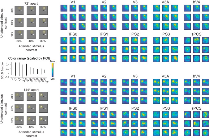
Fig. 4.
Reconstructed spatial maps index stimulus salience and relevance. Image reconstructions are computed for each stimulus separation condition (grouped rows) and each stimulus contrast pair (entries within each 3 × 3 group of reconstructions). Within each group of reconstructions, the diagonals (top left to bottom right) are trials with matched contrasts and are useful for visualizing the qualitative effect of behavioral relevance (spatial attention) on map profiles. Even in V1, locations near attended stimuli are represented more strongly than those near unattended stimuli. Going from top to bottom (unattended stimulus) and left to right (attended stimulus) within each group of reconstructions steps through increasing stimulus contrast levels and can be used to infer the sensitivity of each visual field map to visual salience. Color mapping scaled within each region of interest (ROI) independently (see inset, left center). Range subtended by vertical line for each ROI indicates color range (see color bar) for that ROI. BOLD, blood oxygen level dependent; V1–hV4, visual cortex areas; IPS0–IPS3, intraparietal sulcus regions; sPCS, superior precentral sulcus.

