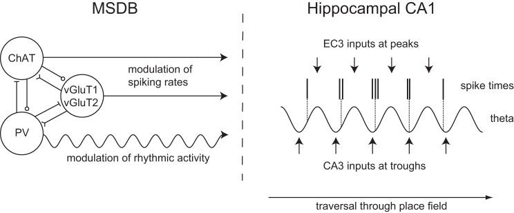
Fig. 4.
Regulation of hippocampal theta rhythm and spike-phase coupling by medial septal inputs. Left: parvalbumin-positive GABAergic, ChAT-positive cholinergic, and vGluT1- or vGluT2-positive glutamatergic MSDB neurons interact via local intraseptal connections and project to the hippocampus. PV-positive neurons can be activated by local cholinergic and glutamatergic inputs. Right: rhythmic inputs from GABAergic MSDB neurons pace hippocampal theta rhythmic activity via disinhibition. Inputs from EC3 and CA3 onto CA1 arrive at the peak and trough of the extracellular theta cycle measured at the pyramidal cell layer, respectively. The balance between EC3 and CA3 relative input strengths might determine the precise spike timing as suggested by different models for phase precession and models of encoding and retrieval. Changes in this balance during the traversal of the place field result in spikes occurring at progressively earlier phases of the theta cycle, i.e., phase precession. ChAT, choline-acetyltransferase; EC3, entorhinal cortex layer 3; MSDB, medial septum and diagonal band of Broca; PV, parvalbumin; vGluT, vesicular glutamate transporter.

