Figure 1.
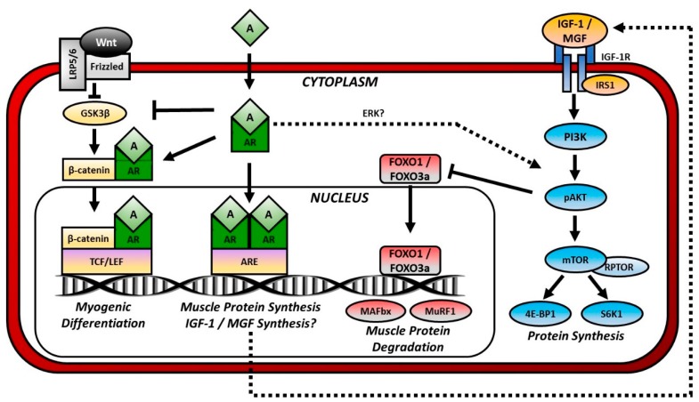
Androgen-mediated Anabolic and Anticatabolic Signaling Pathways in Muscle. Anabolic signaling: Androgens (A) pass through the plasma membrane and bind to cytosolic androgen receptors (AR). Dimerized and phosphorylated ARs pass through the nuclear membrane and bind to a region of the DNA termed the androgen response element (ARE), thereby initiating protein synthesis. Ligand-bound ARs may also enhance Wnt signaling as follows. Wnt binds to Frizzled and in turn disheveled (not shown). Disheveled inhibits the activity of glycogen synthase kinase-3β (GSK3β), which phosphorylates β-catenin and marks it for degradation. When GSK3β is inhibited, β-catenin accumulates and enters the nucleus where it binds to a region of the DNA termed the T-cell factor/lymphoid enhancer factor (TCF/LEF) that regulates genes involved in myogenic differentiation. Ligand-bound ARs enhance Wnt signaling by inhibiting GSK3β and attaching to β-catenin for nuclear shuttling. Androgens may also indirectly stimulate protein synthesis by activating the phosphatidylinositol-3 kinase (PI3K)/Akt signaling through actions of Erk or by promoting synthesis of insulin-like growth factor (IGF)-1 or mechano growth factor (MGF). IGF-1 and MGF bind cell-surface IGF-1 receptors (IGF-1R) and activate PI3K/Akt signaling. Anticatabolic signaling: Activation of PI3K/Akt signaling inhibits the transcription factor forkhead box O (FOXO). FOXO1 and FOXO3a activate muscle atrophy F-box (MAFbx or atrogin-1) and muscle ring finger-1 (MuRF1), and E3 ubiquitin ligases that prepare proteins for proteasome degradation.

