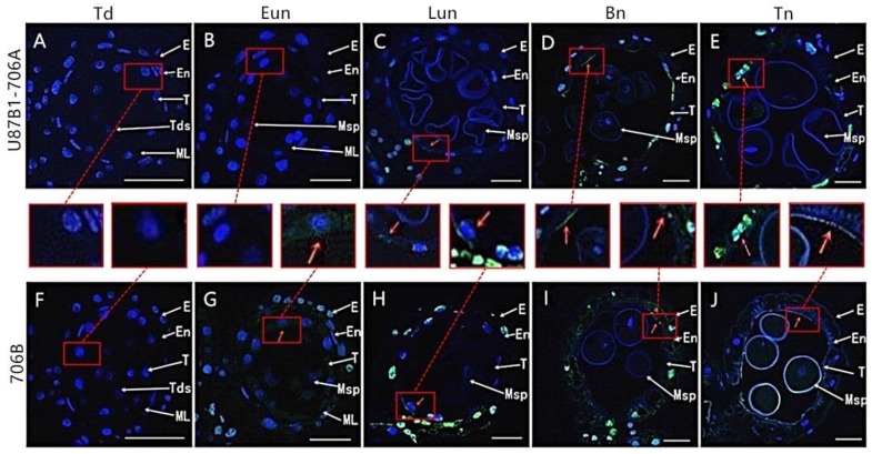
Figure 4.
TUNEL assays to detect anther tapetum PCD in U87B1-706A (A–E) and 706B (F–J) during different developmental stages. (A,F) Td, tetrad stage; (B,G) Eun, early uninucleate stage; (C,H) Lun, late uninucleate stage; (D,I) Bn, binucleate stage; and (E,J) Tn, trinucleate stage. E, epidermis; En, endothecium; ML, middle layer; T, tapetum; Tds, tetrads; Msp, microspores. The green fluorescence denoted by the red arrows indicates nuclei with TUNEL-positive staining. To make green fluorescence visible, the red square shows enlarged example. Scale bars are 50 μm in (A–J).

