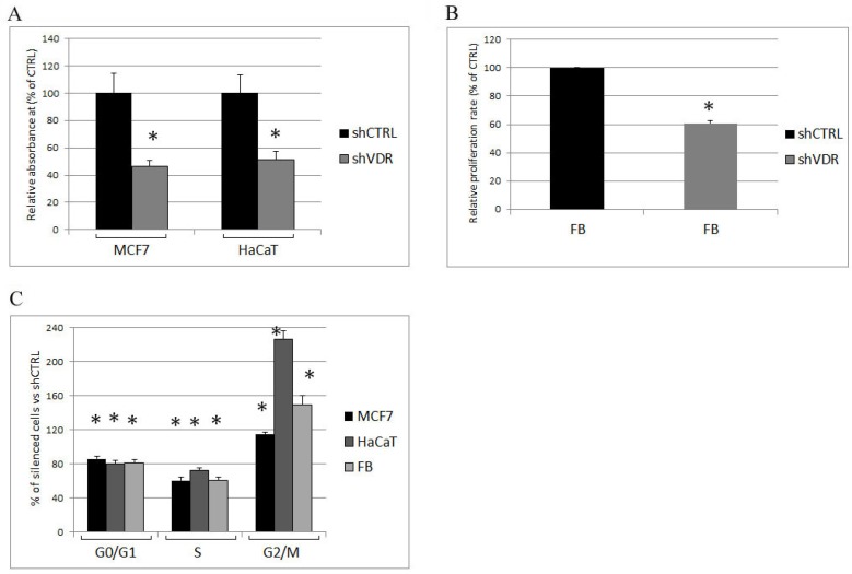
Figure 2.
Analysis of cell proliferation in silenced cells. One week after infection, the control (shCTRL) and VDR knockdown cells (shVDR) were seeded and assayed for (A) proliferation rate, measured by crystal violet staining or (B) BrdU incorporation; (C) The cell cycle of MCF7, HaCaT, and fibroblasts (Fb) was evaluated by cytofluorimetry, and the distribution of the silenced cells throughout the cell cycle was expressed as percentage of the shCTRL cells in the same phase. The data are expressed as the means ± SD of three independent experiments; * p < 0.05 compared to the control.

