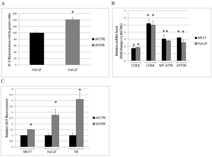
Figure 3.
The silencing of VDR induces mitochondrial respiration and enhances the production of ROS. The metabolic assays and the extraction of mRNA were carried out one week after silencing the cells with shRNA control (shCTRL) or VDR shRNA (shVDR). (A) The mitochondrial respiratory activity was assessed in HaCaT cells by cytofluorimetric evaluation of the mitochondrial dye JC-1, and (B) the expression of the respiratory chain complexes was analyzed by real-time PCR. The values plotted on the graph represent the fold change in transcript expression in silenced versus control cells and are displayed as the means ± SD of three independent experiments; (C) reactive oxygen species (ROS) production was measured and expressed relative to control cells. The data represent the means ± SD of three independent experiments; * p < 0.05 compared to the control.

