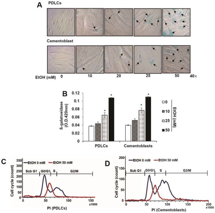
Figure 2.
Effect of ethyl alcohol (EtOH) on characterization of cellular senescence by senescence-associated β-galactosidase (β-gal) staining (A), β-gal activity (B), cell cycle analysis (C,D) and expression of senescence-associated proteins (E) in periodontal ligament cells (PDLCs) and cementoblasts. Cells are incubated with indicated concentration of EtOH for 3 days (A–E); (A,B) SA-β-Gal activity was evaluated using a staining kit. Cell cycle and protein analysis were assessed by flow cytometry (C,D) and Western blot (E), respectively. Flow-cytometric frequency histograms of progenitors stained with propidium iodide (PI) for DNA content. These data are representative of three independent experiments. * statistically significant difference compared to the control groups (p < 0.05). Arrows in Figure 2A represent β-gal (+) cells.

