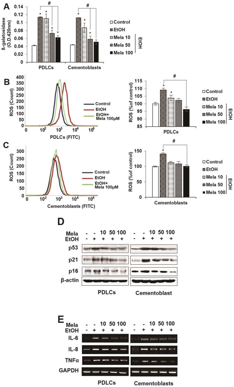
Figure 4.
Effect of melatonin on EtOH-induced cellular senescence in PDLCs and cementoblasts. Cells are incubated with indicated concentration of melatonin (μM) and EtOH (25 mM) for 3 days (A–C). Senescence was examined by β-gal activity (A), ROS production (B,C) and expression of senescence-associated proteins (D) and mRNAs (E). These data are representative of three independent experiments. * statistically significant difference compared to the control groups (p < 0.05). # statistically significant difference in each group.

