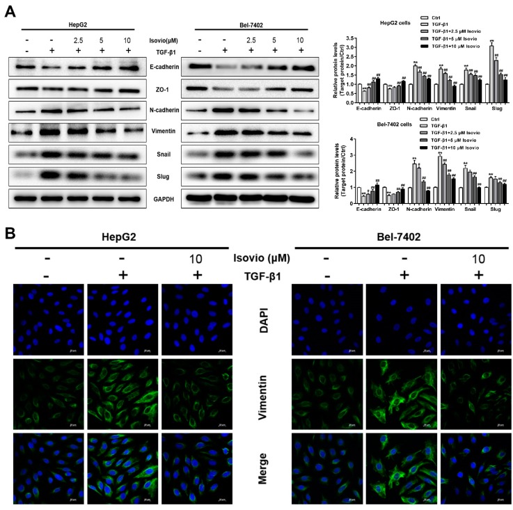
Figure 5.
Effects of isoviolanthin on TGF-β1-induced epithelial–mesenchymal transition (EMT) in HCC cells. (A) HepG2 and Bel-7402 cells were treated with 10 ng/mL TGF-β1 for 48 h, during which the indicated concentrations of isoviolanthin were added for the last 24 h. The expression of the EMT markers E-cadherin, ZO-1, N-cadherin, vimentin, Snail, and Slug was assessed by Western blotting. GAPDH was used as a loading control; (B) E-cadherin and vimentin expression in HCC cells was determined by confocal microscopy. Green fluorescence indicates E-cadherin- and vimentin-positive expression, and blue fluorescence indicates 4’,6-diamidino-2-phenylindole (DAPI)-labeled nuclei. Scale bars: 20 µm. Representative images and bar graphs (mean ± SD) are shown, n = 3. ** p < 0.01 versus the control group; # p < 0.05, ## p < 0.01 versus the TGF-β1 group.

