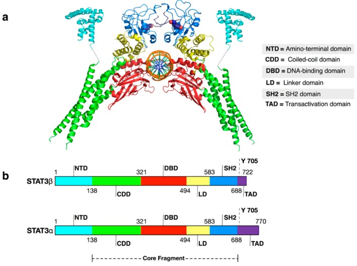
Figure 1.
(a) Cartoon representation of USTAT3β: DNA structure (PDB ID 4ZIA for the N-termini and 4E68 for the remaining structure). Color keys: cyan = amino-terminal domain; green = coiled-coil domain; red = DNA-binding domain; yellow = linker domain; blue = SH2 domain; violet = transactivation domain; orange = DNA. Tyrosine 705 residues are shown as spheres. In the lower part of the picture, a scheme of STATs domain division is reported; (b) Schemes of STAT3α and STAT3β domain division. The dashed line represents the core fragment of the STATs domain (inspired by a scheme presented by Chen et al. [26] for STAT1).

