Figure 3.
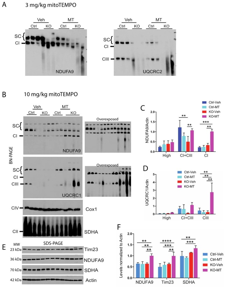
Analysis of mitochondrial supercomplexes in piriform cortex of the neuron-specific RISP KO mice treated with mitoTEMPO. Mitochondrial fraction from piriform cortex were obtained from control and RISP KO mice treated with either vehicle or with (A) low dose of 3 mg/kg/day or (B) high dose of 10 mg/kg/day of mitoTEMPO (MT). Mice were treated from weaning to 82–105 days of age. SCs were analyzed by BN-PAGE and western blot to detect SCs (HMW and CI+CIII), CI, CIII, CIV and CII using antibodies against NDUFA9, UQCRC1, Cox1 and SDHA subunits respectively. Low dose did not improved stability of respiratory complexes. Graphs represent the quantification of the levels of NDUFA9 (C) and UQCRC1 (D) normalized to actin in HMW SCs, CI+CIII, free CI or CIII respectively of the blots shown in (B). (E) Steady-state levels of some mitochondrial proteins in piriform cortex of vehicle and MT-treated mice by SDS-PAGE. Molecular weight (MW) for each protein is indicated in the figure. (F) quantification of blots in (E). Quantification of signal in blots was performed by densitometry analysis with ImageJ software. Protein levels were normalized to actin. Graphs represent the mean and standard deviation. Statistical analysis was performed by one-way ANOVA followed by Tukey’s multiple comparison test. (**) p < 0.01; (***) p < 0.001; (****) p < 0.0001 indicates statistical significance, n = 3–4.

