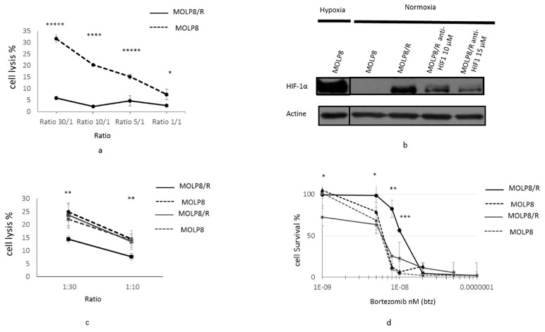
Figure 4.
Resistance of MOLP8/R cell line to the immune system: role of HIF1α. Tumor cell lysis MOLP8 and MOLP8/R by NK92MI cell line at different ratios (tumor cells/NK cells). Tumor cells are incubated for 4 h in contact with NK cells, then stained with carboxyfluorescein succinimidyl ester (CFSE) and Thiazol Orange PRO (TOPRO). The double positivity of tumour cells by both dyes are determined by flow cytometry. Black dotted line is MOLP8, and black line is MOLP8/R (a). Involvement of HIF 1α in resistance to NK cell lysis. Both cell lines MOLP8 and MOLP8/R are treated for 48 h with an anti-HIF1α drug (cryptotanshinone) at two different concentrations, 10 and 15 µM. The validation of the inhibition of HIF1α in MOLP8/R is verified by Western blot (b). Afterwards, treated cell lines MOLP8/R and MOLP8 are incubated for 4 h with NK cells, and the lysis is evaluated by flow cytometry. Black dotted line is MOLP8, and black line is MOLP8/R, grey dotted line is MOLP8 after treatment, and grey line is MOLP8/R (c). Involvement of HIF1α in drug resistance to Btz, Molp8, and MOLP8/R is treated and co-treated for 48 h with Btz, or Btz plus cryptotanshinone at 10 µM, and the cytotoxicities are calculated. Black dotted line is MOLP8, and black line is MOLP8/R, and after treatment, grey dotted line is MOLP8, and grey line is MOLP8/R (d). * p < 0.05, ** p < 0.01, *** p < 0.001, **** p < 0.0001 and ***** p < 0.00001.

