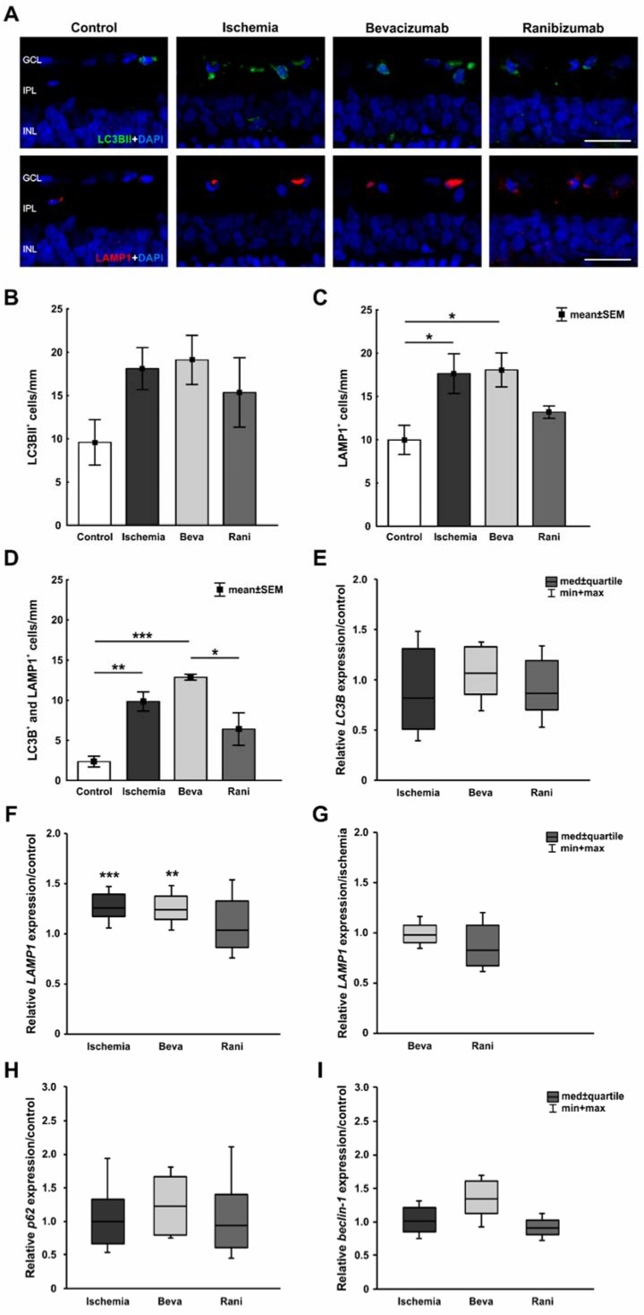
Figure 3.
(A) Early autophagy was labelled with anti-LC3BII (green), while anti-LAMP1 was used to visualize the late autophagy (red) and DAPI-marked cell nuclei (blue). More autophagocytotic cells were noted after ischemia induction; (B) LC3BII+ cell counts revealed no differences between all ischemic groups (p > 0.05), when compared to controls, although the number of positive cells was a little bit higher in those groups; (C) Regarding LAMP1 staining, significantly more LAMP1+ autophagocytotic cells were detected in ischemic (p = 0.04) and bevacizumab-treated (p = 0.03) eyes, when compared to controls. The retinae treated with ranibizumab displayed fewer LAMP1+ cells, no significant difference could be observed in relation to control eyes (p = 0.6); (D) Equally, evaluation of colocalized LC3BII+ and LAMP1+ cells revealed significantly increased LC3BII+ and LAMP1+ cell numbers in the ischemic (p = 0.007) and bevacizumab-treated groups (p < 0.001), but not in ranibizumab-treated retinae (p = 0.2); (E) Via qRT-PCR no differences in relative LC3B mRNA expression were noted when compared the untreated (p = 0.46) and treated ischemic groups (beva: p = 0.625; rani: p = 0.38) to the control group; (F) A significant up-regulation of LAMP1 mRNA levels could be measured in the untreated ischemic (p < 0.001) and bevacizumab-treated groups (p = 0.007) in relation to control retinae. No differences in relative LAMP1 mRNA expression were seen between the control and ranibizumab-treated retinae (p = 0.769); (G) In comparison to the ischemic group, a slight trend to less LAMP1 mRNA expression was noted in retinae treated with ranibizumab (p = 0.13); (H) Regarding p62 mRNA expression, no effect on the expression level was observed between control retinae and all ischemic groups (p > 0.05); (I) The relative beclin-1 mRNA expression levels in the ischemic (p = 0.888), bevacizumab (p = 0.086), and ranibizumab-treated eyes (p = 0.178) were similar to the control group. *: p < 0.05; **: p < 0.001; ***: p < 0.001. Abbreviations: GCL: ganglion cell layer, IPL: inner plexiform layer, INL: inner nuclear layer, Beva: bevacizumab, Rani: ranibizumab. Scale bar: 20 µm. Immunohistology: n = 5–6/group; qRT-PCR: n = 4/group.

