Fig. 5.
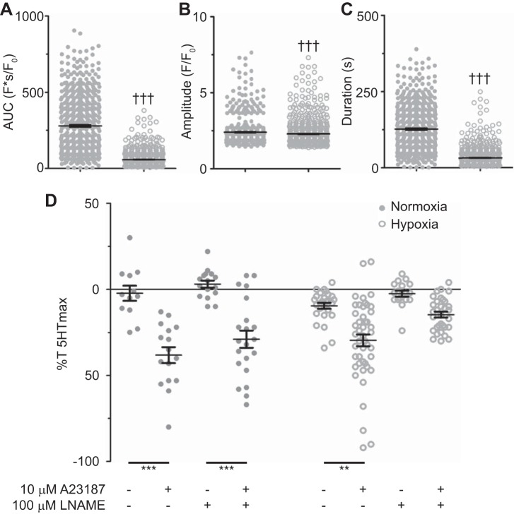
Long-term hypoxia (LTH) modifies A-23187-mediated relaxation and Ca2+ signals. Area under the curve (A), duration (B), and amplitude of the Ca2+ events (C) for regions of interest that were automatically detected in normoxic (n = 5/553) and LTH (n = 7/563) sheep arterial myocytes. D: relaxation relative to the contraction induced by 1 μM serotonin (5-HT) in normoxic (n = 5/71) and LTH (n = 8/100) sheep pulmonary arteries. Individual data points are shown for normoxic (closed) and LTH (open) recordings, and bars indicate means ± SE †††P < 0.001, statistically significant effect based on a Mann Whitney U-test. **P < 0.01 and ***P < 0.001, statistically significant effect based on a Kruskal-Wallis 1-way ANOVA with a Dunns multiple-comparison test.

