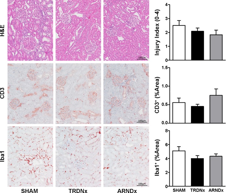
Fig. 6.
Histological assessment of renal injury and immune cell infiltration. In a subset of animals in each group (n = 6/group), renal injury, T cell infiltration, and macrophage infiltration were assessed by histological analysis. Hematoxylin and eosin (H&E)-stained tissues were scored by a blinded investigator for injury. Immunohistochemical staining for CD3 and Iba1 was employed to quantify T cell and macrophage density in renal parenchyma, respectively. Together, no effect of total renal denervation (TRDNx) or afferent-selective renal denervation (ARDNx)was detected in any histological measurement. Representative images of cortical tissue are presented at ×10 magnification. Scale bar = 100 µm. Data are presented as means ± SE.

