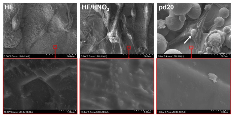
Figure 7.
hMSCs under SEM microscope spreading after 1 day of culture on the polished (HF and HF/HNO3 solutions) and unpolished (45 W laser power, 40 μs exposure time, 20 μm point distance) titanium discs. Red squares indicate area of the samples which are magnified to show sample topography and are depicted on images below.

