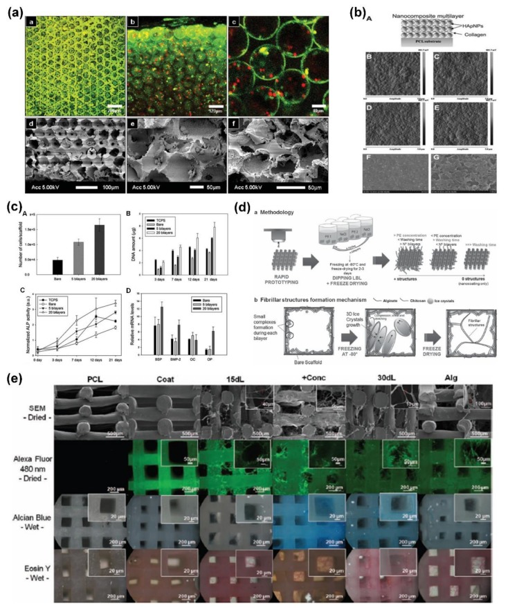
Figure 5.
(a) Confocal images of inverted colloidal crystal scaffolds cultured with thymic epithelial cells and monocyte cells; (b) illustration of LbL multilayer nanocomposite coating with hydroxyapatite and collagen on substrates, and the AFM images of multilayers with different numbers of bilayers; (c) hMSCs adhesion and their quantification of DNA amounts to bare and coated scaffolds, and alkaline phosphatase activity and relative mRNA expression during the culture of hMSCs on various substrates; (d) steps and mechanism for developing the hierarchical and hybrid 3D scaffolds and (e) representative images of the structures of these scaffolds. Reprinted from [49,92,94] with permissions from Wiley-VCH and Royal Society of Chemistry.

